January 3, 2005
Femtosecond laser technique opens new opportunities
for research on nerve regeneration
By Tim Stephens
In a breakthrough for research on nerve regeneration, a team
of UCSC and Stanford scientists has reported using femtosecond
laser pulses to precisely cut individual axons of nerves in
the roundworm Caenorhabditis elegans, one of the most
versatile and widely used experimental organisms for genetic
and biomedical research.
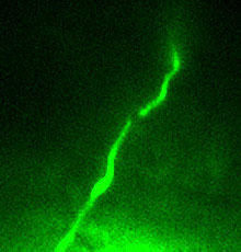
This nerve axon was cut using
femtosecond laser nanosurgery.
Photo: Yanik et al.
|
The nerves severed by this precision technique regrew within
24 hours, often with complete recovery of function.
The project was a collaboration between applied physics researchers
at Stanford University led by Adela Ben-Yakar and biologists
at UCSC led by Yishi Jin and Andrew Chisholm.
The team's findings give researchers an experimental system
in which they will be able to investigate in great detail the
genetic and molecular factors that control whether or not damaged
nerves can regrow, said Chisholm, an associate professor of
molecular, cell, and developmental biology.
"This technique will enable us to find the genes that
are important in allowing an axon to regenerate. In the worm,
we can do systematic screening of large numbers of genes, and
of drugs and other small molecules as well, to ask how they
affect the process of regeneration," Chisholm said.
The researchers reported their findings in a paper published
in the December 16 issue of the journal Nature. The first
author of the paper is Mehmet Fatih Yanik, a Stanford graduate
student in applied physics, who worked with Ben-Yakar in her
femtosecond laser nanosurgery project.
Ben-Yakar initiated the project two years ago at Stanford and
is continuing it as an assistant professor of mechanical engineering
at the University of Texas at Austin. The other coauthors include
Jin, a professor of molecular, cell, and developmental biology
at UCSC and a Howard Hughes Medical Institute investigator;
Hulusi Cinar, a postdoctoral researcher in Jin's lab; and Hediye
Nese Cinar, a postdoc in Chisholm's lab.
The Cinars met Yanik through personal connections and initially
discussed possible collaboration about a year ago. Yanik later
told them about the femtosecond laser and how other researchers
had begun using it in biological systems to surgically destroy
extremely tiny structures.
"When Yanik described to me what this instrument can do,
I immediately thought of my work on the nervous system of C.
elegans and came up with a nerve regeneration experiment
we could do together, and I designed the experiments,"
said Hulusi Cinar.
Yanik performed the nanosurgery procedure at Stanford. The
technique uses extremely short pulses of intense laser light
to focus energy in a very small volume. When properly focused,
the energy delivered by the laser pulses breaks down chemical
bonds at the targeted site, vaporizing the tissue within a tiny
volume without causing side effects such as heating of surrounding
tissue, Yanik said.
The duration of the laser pulses used in the study was 200
femtoseconds (a femtosecond is a millionth of a billionth of
a second), and the pulses were delivered at a rate of one thousand
per second. The delicate axons severed by the procedure, with
no apparent damage to surrounding tissue, were on average just
0.3 microns, or 300 nanometers, in diameter (a nanometer is
one billionth of a meter).
"I am very excited about the merging of this new technology
into biological research. I didn't know anything about femtosecond
laser technology until the physicists explained it to me,"
said Jin, who has spent years investigating the development
of the worm's nervous system. "Now there is a lot to do--it
has opened up the potential to address questions that we have
never been able to address before."
In their experiments, the researchers cut a nerve that runs
from one side of the worm's body to the other. The nerve inhibits
contraction of muscles on one side of the body while the muscles
on the other side are contracting. It functions during the alternating
contractions of muscles on either side of the body that enable
the worm to wiggle backward with a smooth wavelike motion. Hulusi
Cinar and Jin have been studying so-called "shrinker"
mutants that lack this ability due to a genetic defect.
"A normal worm, when you touch its head, will move backward
in a smooth motion. In the shrinker mutants, the muscles on
both sides contract simultaneously, so they don't move back,"
Cinar said. "So these neurons were a good target for surgery
because we knew that when they are knocked out you get a well-defined
behavioral effect, and it's straightforward to see if their
function has been recovered through regeneration."
Nese Cinar designed a movement assay to evaluate the behavioral
effects in the worms and evaluated the worms in the experiments
in a "blinded" manner, not knowing which ones had
received the surgery.
"Without such functional assays, any anatomically observed
regeneration could be explained in various ways, such as bleaching
and recovery of the marker protein used to label the neurons,"
she said.
The nanosurgery, performed on anesthetized juvenile worms,
could be completed in about 10 minutes per worm once the equipment
was set up.
Although regeneration of peripheral nerves is nothing new,
Chisholm said he was still surprised by the rapid recovery of
function in the worms.
Most of the severed axons regrew within 12 to 24 hours after
the laser surgery. Preliminary observations indicated that after
an axon is cut, the nerve cell sprouts a new axon from the severed
end that regrows to reach the target muscle. In some cases,
however, it appears that the two severed ends reattach, Chisholm
said.
"Clearly there is a lot more biology going on here that
we need to explore. This just opens up a lot of exciting things
to study," he said.
C. elegans, an almost microscopic nematode or roundworm,
has become an extremely important system for biomedical research.
Geneticists have identified thousands of genes in the worm that
have counterparts in humans. A relatively simple organism with
a short generation time, reproducing in just three days, it
is easy to study in the laboratory. It has many of the same
basic physiological and anatomical features, such as muscles
and nerves, found in more complex animals. And it is even transparent,
making it easy to see things like nerves inside its body. The
researchers who pioneered the use of C. elegans for biomedical
research received the 2002 Nobel Prize for physiology and medicine.
Now this excellent model system for biomedical research is
available for studying nerve regeneration. One of the fundamental
questions researchers want to answer is why nerve damage in
the central nervous system--the brain and spinal cord--is usually
permanent.
"In humans, peripheral nerves will regrow, but in the
central nervous system the regrowth of axons is inhibited by
the local environment. That's why spinal cord injuries are so
serious. We want to find out why a severed axon will regrow
in some situations and not in others," Chisholm said.
Return to Front Page